
Page 27 - VOL29_N_75_2017
P. 27
Case report 2
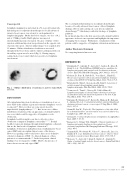
Lymphatic mapping was performed in a 54-year-old woman who had undergone neoadjuvant chemotherapy for locally advanced invasive breast cancer, was referred to our department for lymphoscintigraphy. On the day before surgery, one dose of 0.2 mL of 37 MBq (1 mCi) 99mTc-phytate was injected interadermally in periareolar region. Because of failure of first injection, additional injection was performed on the opposite side of periareolar region. Anterior planar images were acquired after 15 minutes. Diffuse distribution of radiotracer was noted throughout the breast tissue and two faint focal hyperactivities in the axillary region was also noted (Fig. 2). During surgery, sentinel nodes were found, which were positive for lymphatic involvement.
Fig. 2 - Diffuse distribution of radiotracer and two faint SLNs are seen.
DISCUSSION
After intradermal injection of radiotracer, visualization of one or more SLNs in the axillary region and sometimes lymphatic vessels is expected [1]. However, there are some reports describe unpredictable lymph pathways [2, 4, 5]. But, in our cases, diffuse distribution of radiotracer like an appearance of dermal backflow was seen, which could be suggestive of lymphatic vessels obstruction.
Lymphatic backflow might occur due to defective valves, obstruction or impaired lymphatic contractility[4, 6, 7]. Dermal backflow represents drainage through dermal lymphatic collaterals that are the final result of lymphatic obstruction [8].
Lymphatic obstruction is pathophysiology of inflammatory breast cancer (IBC) and can explain clinical course of the disease. However, regarding controversial issues, there are no definitive criteria for diagnosis of IBC. So, delayed diagnosis of these patients is a common mistake[9]. In the first patient, clinical findings were not suggestive of IBC. Maybe, the presence of denser breast was an early sign of IBC.
The second patient had undergone neoadjuant chemotherapy because of locally advanced breast cancer. Altered lymphatic drainage, fibrosis and scarring can occur after neoadjuvant chemotherapy [10], which may resulted in blockage of lymphatic pathways.
In our knowledge, this is the first report describe dermal backflow appearance in breast cancer patients and highlights the importance of imaging before surgery. Furthermore, visualization of this pattern could be suggestive of lymphatic obstruction and invasion.
Author Disclosure Statement
No competing financial interests exist.
REFERENCES
11. Giammarile F., Alazraki N., Aarsvold J., Audisio R., Glass E., Grant S. et al.: The EANM and SNMMI practice guideline for lymphoscintigraphy and sentinel node localization in breast cancer. Eur J Nucl Med Mol Imaging, 2013; 40(12): 1932-47.
12. Groheux D., Ferré R., Rubello D., Vercellino L., Hindié E.: Breast cancer patient with an uncommon lymphatic drainage evidenced by SPECT/CT. Clin Nucl Med, 2014; 39(2): 176-9.
13. Krynyckyi B., Shim J., Goyenechea M., Kim C.: Layering of activity around a breast implant capsule during lymphoscintigraphy. Clin Nucl Med, 2004; 29(9): 598-9.
14. Vermeeren L., Tanis P., Nieweg O., Valdés Olmos R.: Lymphoscintigraphy of a breast tumor showing focal tracer accumulation along the falciform ligament of the liver. Clin Nucl Med, 2010; 35(3): 168-9.
15. Barranger E., Montravers F., Kerrou K., Marpeau O., Raileanu I., Antoine M. et al.: Contralateral axillary sentinel lymph node drainage in breast cancer: a case report. J Surg Oncol, 2004; 86(3): 167-9.
16. Suami H., Pan W., Taylor G.: The lymphatics of the skin filled by a dermal backflow: an observation in a scarred cadaver leg. Lymphology, 2007; 40: 122-6.
17. Zawieja D.: Contractile physiology of lymphatics. Lymphat Res Biol, 2009; 7: 87-96.
18. Karaçavus S., Yilmaz Y.K., Ekim H.: Clinical significance of lymphoscintigraphy findings in the evaluation of lower extremity lymphedema. Mol Imaging Radionucl Therapy, 2015; 24(2): 80-4.
19. Yamauchi H., Woodward W., Valero V., Alvarez R., Lucci A., Buchholz T. et al.: Inflammatory breast cancer: what we know and what we need to learn. Oncologist, 2012; 17(7): 891-9.
10. Moncayo V., Aarsvold J., Grant S., Bartley S., Alazraki N.: Status of sentinel lymph node for breast cancer. Semin Nucl Med, 2013; 43(4): 281-93.
THE EUROPEAN JOURNAL OF LYMPHOLOGY - Vol. XXIX - Nr. 75 - 2017
23
