
Page 19 - VOL29_N_75_2017
P. 19
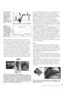
Graph 1: Example of a recorded pressure curve: base line period before distal paw immersion (A), immersion paw step (B), post-immersion paw period (C) and pressure difference (D).
Graph 2: Plethysmometer calibration curve (linear regression). Statistical software: GraphPad Prism 4, Goodness of the fit (Sy.x = 0.006836) and repeated pressure measures (8 times) of 12 known volumes.
In the experimental group, the rats underwent a combined left superficial dissection of the brachial nodes (Fig. 2b) and a deep dissection of the axillary nodes (Fig. 2c) by an innovative posterior surgical approach (Fig. 2a) (Pastouret et al., 2016). The technique concerns a skin incision of 1 cm length parallel to the posterior border of the triceps brachialis. The space between the interior face of the skin and the subcutaneous aponeurosis was split to assess directly the brachial nodes. Total fat tissue containing the brachial nodes was dissected followed by further dissecting and ligating the vascular pedicles and removal of the brachial nodes. Access to the axillary cavity was performed through an inter- aponeurotic passage between the cutaneous trunci and latissimi dorsi muscles. Hidden in the cavity, the axillary nodes were then delicately searched for, dissected and resected taking care that the elements of the peripheral neurovascular mass remained intact.
At 12 weeks, the ultimate paw volume assessments were carried out under general anaesthesia (same protocol). Then ICG mapping was accomplished before collection of skin samples to detect LFS and after collection of skin samples to detect FLSP. Indirect lymphographies were performed following subcutaneous injection of ICG at the back side of the forefoot of each investigated front paw (0.05 ml, 5 mg/ml, Pulsion Germany). Massage at the injection site was done during 3 minutes for enhancing the absorption of tracer by the initial lymphatics and to help out its evacuation in the lymphatic collector vessels (Gashev et al., 2010; Unno et al., 2008). The ICG mappings were achieved by an artisanal near infrared camera.
The near infrared light source consisted of two parts. A 30 LED light ring (760 nm) was fixed to the camera for a general illumination of the target zone and a 30 LED mobile light module (760 nm) focused an additional illumination on a more specific part of the target. In addition, one long pass filter (Schott RG830 or Schott RG850) was placed in front of the lens of the digital camera which was connected to the near industrial camera. The choice of the filter depended on light environmental conditions. Focusing and zooming the lens could be manually adjusted. These two additional settings solved various disadvantages of the other near infrared cameras available in the medical field.
At the end of the experiment, rats were euthanized by injecting an overdose of Nembutal.
Part 2
Female N.M.R.I. (Naval Medical Research Institute, Bethesda, Maryland, USA) white mice, aged 6 to 8 weeks and weighing 28 to 30 grams, were selected for this experiment. The lymphatic vessel, popliteal and sacral lymph nodes of the posterior paw were used as lymphatic system model (Fig. 3). The investigations were approved by the local Animal Care Committee of the Vrije Universiteit Brussel.
Animal distribution was randomized in three groups of mice: 5 mice in the massage group, 10 animals in the vibration group and 10 mice in the control group. All animals were anaesthetized with urethane (2 mg/g i.p.). Hair on the posterior paws was shaved with electric clippers.
A spectrophotometric technique was chosen for the analysis of lymph node blueness to obtain quantitative values of the lymphatic functions (reabsorption and lymph flow). Having completed lymph node resections, Evans blue dye (EBD) was extravasated from the nodes according to the method used by Greco (Greco et al., 2006).
Fig. 3 - Injection site and visualisation of the lymphatic vessel, popliteal and sacral lymph nodes (coloured by Patent Blue) in the mouse posterior paw.
Fig. 2 - Innovative posterior surgical approach.
(a) Location of the skin incision. (b) The main superficial lymphatic pathway following skin dissection: the white arrow indicates the Evans Blue dye injection site which causes the lymphatic vessels to become visible (grey arrows). The black arrow indicates the brachial nodes in fat tissue. (c) Following cutaneous trunci muscle section (white triangle), the deep axillary nodes are located. The black arrow indicates the axillary node and the white star is positioned over the latissimus dorsi muscle.
THE EUROPEAN JOURNAL OF LYMPHOLOGY - Vol. XXIX - Nr. 75 - 2017
15
