
Page 20 - VOL29_N_75_2017
P. 20
Experimental protocol
Following anaesthesia and shaving, the animals were placed in an identical position on a same platform allowing them to vibrate. Respecting five minutes of rest, an EBD solution (25 mg/ml, 20 μl) was injected subcutaneously at the dorsal side of the foot pad over a period of three minutes. Bilateral injections were carried out in the massage group. The choice of the first foot pad to be injected was randomized. Unilateral injections were performed in both vibration and control groups where the foot pad to be injected was randomized as well.
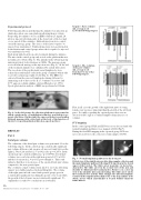
Each group underwent its specific treatment during five minutes. The mice in the control group only rested on the platform that was not induced to vibrate (Fig. 4). The animals in the vibration group underwent whole body vibrations at 30 Hz. The massage group received bilateral massage at the injection sites (Fig. 4). At the end of the treatment animals were euthanized by cranial dislocation. The resections of the popliteal and sacral lymph nodes were realized in a randomized order in the group of animals which only received local massage (right or left) (Fig. 4). The EBD was extracted from the removed lymph nodes during 24 hours by immersing each of these nodes in a 7:3 mixture of acetone and 0.5% aqueous sodium sulphate solution (Harada et al., 1971). Spectrophotometric analysis of EBD was performed at 620 nm.
Fig. 4 - In the left picture the vibration platform is represented in which a single motor of Andullation technology was built in to ge- nerate vibrations. On the right, the same researcher is performing bilateral massage with cotton buds at the level of the injection of the dye by respecting identical direction, speed and force.
RESUL TS
Part 1
Distal paw volumes
The evaluation of the distal paw volumes was performed 12 weeks following surgery. In the control group, a statistically significant paw volume difference (ml, mean ± sd) was not found between the right (2.59 ± 0.29) and left (2.37 ± 0.42) sides (p = 0.2073). In the operated group, a statistical difference (p = 0.0350) of front paw volumes was seen between the right non-operated (2.53 ± 0,26) and the left operated (2.36 ± 0,31) paws (Graph 3). This result indicates that there is no increase of the operated front paw volume compared to the non-operated paw.
The evolutions of paw volumes from day 0 to 12 weeks following surgery were further analysed in detail (Graph 4). The volume
of the right paws in both control and operated groups grew in
a statistically significant way during the period of 12 weeks while the growth of the left paws was not statistically significant. To explain these results, two conclusions are forwarded.
THE EUROPEAN JOURNAL OF LYMPHOLOGY - Vol. XXIX - Nr. 75 - 2017
Graph 3 - Paws volumes (ml, average ± sd)
12 weeks following surgery.
16
Graph 4 - The evolution of the paw volumes
(ml, average ± sd)
from day 0 to 12 weeks following surgery.
First, in all cases the growth of the right front paws of young females rats was more important than the growth of the left front paws. Secondly, regarding the operated paws, there was no detected visible sign of secondary lymphoedema (increase of volume).
ICG mapping
In the control group, FSLP and LFS were never detected and only normal lymphatic pathways were mapped (20/20) (Fig 5).
During the first ICG mapping in the operated group, LFS was never detected before the collection of skin samples (60/60).
Fig. 5 - Normal lymphatic pathways in the front paw.
Global view of the right front paw after skin samples collection and the main lymphatic pathway following ICG injection in the dorsal aspect of the forefoot. The white arrow in picture (a) shows bra- chial nodes. The white arrow in the zoomed view of the arm and shoulder (b) illustrates the brachial nodes. Picture (c) is a close-up view of the shoulder after arm stretching. The white arrow shows the brachial nodes and the white triangle the efferent lymphatic vessel from the superficial brachial nodes to the deep axillary node (black arrow) which anatomically is located behind cutaneous trunci muscle.
